Understanding how does a medical ventilator work is essential for patients, families, and healthcare professionals in 2026. These machines support breathing when the lungs cannot move enough air independently. They do not simply “push oxygen” into the body. Instead, a ventilator delivers carefully measured breaths through a mask or breathing tube. It controls airflow, pressure, volume, oxygen concentration, and breathing timing.
Modern ventilators use sensors to monitor each breath. A screen may show airway pressure, delivered volume, respiratory rate, and oxygen levels. Clinicians adjust these settings according to lung condition, blood-gas results, patient comfort, and treatment goals. For example, a patient with severe lung inflammation may need gentle pressures to reduce additional injury. Another patient may need stronger assistance while recovering from anesthesia or muscle weakness. Small changes matter.
The technology is advanced, but it is not independent care. Nurses and respiratory therapists continually assess the patient, tubing, alarms, and skin around the mask. Doctors also review clinical evidence and manufacturer guidance before changing settings. Trusted standards from organizations such as the FDA, WHO, and professional respiratory-care societies help shape safe practice. Still, no ventilator decision fits every patient. That is where explanations can become too simple. This article explores the breathing cycle, pressure support, oxygen delivery, monitoring, alarms, and newer automated features used in 2026. It also considers practical limitations, because a ventilator supports failing physiology but does not cure every underlying disease.
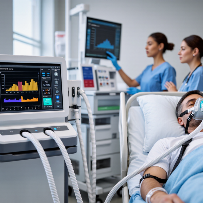
A medical ventilator is a machine that supports breathing when the lungs cannot move enough air. It pushes a controlled mixture of oxygen and air through a mask or breathing tube. Sensors measure pressure, airflow, and the patient’s response. The device then adjusts support according to programmed clinical settings.
Ventilators may be used during surgery, severe pneumonia, acute respiratory distress, or serious muscle weakness. Some patients need noninvasive ventilation through a tight mask. Others require an invasive tube placed into the airway. The machine can deliver breaths, assist the patient’s own effort, or maintain pressure between breaths. It does not cure the underlying illness. That part requires medical treatment.
The difficult part is choosing the right support. More pressure is not automatically better. Excessive pressure may injure fragile lung tissue, while insufficient support can increase exhaustion. Clinicians watch oxygen levels, blood gases, chest movement, alertness, and alarm patterns. Human judgment still matters.
Tips: Keep the mask sealed, but report pain or air leaks. Never change settings yourself. Ask why each setting is being used. A calm bedside explanation can reduce fear. One detail is easy to miss: comfort affects cooperation, and cooperation can affect breathing.
A medical ventilator moves gas through a controlled breathing circuit. A flow sensor measures each breath, while pressure sensors track resistance and lung stiffness. The clinician sets inspiratory flow, respiratory rate, tidal volume, and expiratory pressure. Software then adjusts valves within milliseconds.
Oxygen delivery is more than turning a dial. The ventilator blends air and oxygen to reach the prescribed fraction of inspired oxygen, or FiO₂. Pulse oximetry and arterial blood gases help assess the result, but neither gives the complete picture. The ARDS Network trial found that 6 mL per kilogram of predicted body weight reduced mortality compared with traditional larger breaths in acute respiratory distress syndrome. It also limited plateau pressure below 30 cmH₂O, reducing excessive lung stress.
Pressure matters.
Positive end-expiratory pressure, or PEEP, keeps vulnerable air sacs from collapsing between breaths. Too little may worsen oxygenation; too much can impair blood return to the heart. The 2024 Global Initiative for Chronic Obstructive Lung Disease report emphasizes individualized oxygen targets for patients at risk of carbon dioxide retention. This is why a fixed oxygen setting can be unsafe.
In practice, alarms may reveal blocked tubing, secretions, leaks, or patient-ventilator mismatch. The machine detects these problems, but it cannot interpret every clinical cause. That limitation matters. A rising pressure waveform may reflect bronchospasm, coughing, or a poorly positioned tube. Clinicians must connect the numbers with bedside observation, repeated assessments, and validated protocols.
Core operating parameters, control mechanisms, and commonly used clinical settings
| System Dimension | What the Ventilator Controls or Measures | Typical Clinical Value or Range | How It Affects Airflow and Oxygen Delivery | Safety or Clinical Consideration |
|---|---|---|---|---|
| Gas Supply | Compressed air and oxygen enter through separate high-pressure inlet ports and are blended by proportional valves. | Air: approximately 21% oxygen Oxygen supply: commonly up to 100% oxygen |
The blended gas passes through the inspiratory circuit at a programmed flow, pressure, or volume. | The delivered oxygen concentration depends on the selected oxygen fraction and the accuracy of the gas blender. |
| Inspired Oxygen Fraction (FiO₂) | The percentage of oxygen in the gas delivered to the patient. | 21%–100% | Increasing FiO₂ raises the available oxygen concentration in the alveoli and can improve blood oxygenation. | The lowest FiO₂ that achieves the prescribed oxygen target is generally preferred to limit unnecessary oxygen exposure. |
| Tidal Volume (Vt) | The volume of gas delivered during each mandatory or assisted breath in volume-targeted ventilation. | Often 4–8 mL/kg of predicted body weight; approximately 6 mL/kg is commonly used in lung-protective ventilation for acute respiratory distress syndrome. | A larger Vt delivers more gas per breath, while a smaller Vt reduces stretch on the lungs. | Vt is based on predicted body weight rather than actual body weight. Clinicians also monitor plateau pressure and driving pressure. |
| Respiratory Rate | The number of mandatory breaths delivered per minute; patient-triggered breaths may occur in addition. | Common adjustable range: about 4–60 breaths/min | Increasing the rate generally increases minute ventilation and carbon dioxide removal, provided adequate expiratory time remains. | An excessive rate can shorten exhalation and contribute to air trapping or dynamic hyperinflation. |
| Minute Ventilation | Total expired or delivered gas volume per minute, calculated approximately as tidal volume multiplied by respiratory rate. | Formula: V̇E = Vt × respiratory rate | Minute ventilation is a major determinant of carbon dioxide clearance. | Effective alveolar ventilation is lower than minute ventilation because some gas occupies anatomical and physiological dead space. |
| Inspiratory Flow | The speed and pattern at which gas enters the patient circuit. | Common adult settings: approximately 20–100 L/min; some systems allow higher peak flows. | Higher flow reaches the set volume faster and may improve comfort during high respiratory demand. Flow pattern also changes inspiratory pressure and timing. | Flow must be coordinated with patient effort to reduce dyssynchrony and avoid unnecessarily high airway pressures. |
| Inspiratory Time and I:E Ratio | The duration of inspiration compared with expiration. | Common starting I:E ratio: about 1:2; longer expiratory times may be used in obstructive disease. | Longer inspiration can improve mean airway pressure, while longer expiration helps reduce air trapping. | The correct timing depends on lung mechanics, respiratory rate, patient effort, and the presence of obstructive disease. |
| Positive End-Expiratory Pressure (PEEP) | Pressure maintained in the airway at the end of expiration. | Often 5–20 cmH₂O in adult critical care; individualized according to oxygenation and lung mechanics. | PEEP helps prevent alveolar collapse and can improve functional residual capacity and oxygenation. | Excessive PEEP may reduce venous return, impair circulation, or overdistend relatively healthy lung regions. |
| Pressure-Controlled Ventilation | The ventilator controls inspiratory pressure and time while the delivered volume varies with compliance and resistance. | Inspiratory pressure is commonly set above PEEP; the exact value is patient-specific. | Flow is usually decelerating: it starts high and decreases as the target pressure is reached. | Delivered tidal volume must be monitored because it can change when lung compliance or airway resistance changes. |
| Volume-Controlled Ventilation | The ventilator targets a set tidal volume and adjusts flow to deliver that volume. | Typical adult Vt: approximately 4–8 mL/kg predicted body weight. | The set volume is maintained, but airway pressure may rise when compliance worsens or resistance increases. | Peak and plateau pressures should be monitored to identify excessive airway or alveolar stress. |
| Pressure Support | Additional inspiratory pressure provided for spontaneous breaths, commonly during assisted or weaning modes. | Often approximately 5–20 cmH₂O above PEEP, adjusted to patient effort and target volume. | Pressure support reduces the work required to overcome airway resistance and circuit load. | Too much support can produce excessive tidal volumes; too little may result in fatigue or inadequate ventilation. |
| Trigger Sensitivity | The pressure or flow change required for the ventilator to recognize a patient’s inspiratory effort. | Common flow-trigger settings: approximately 1–5 L/min; pressure-trigger settings may be around −0.5 to −2 cmH₂O. | A sensitive trigger allows rapid assistance after patient effort begins. | If set too sensitively, leaks or circuit movement may cause auto-triggering; if set too low, the patient may experience excessive trigger work. |
| Cycling-Off Criteria | The condition that ends inspiration and switches the ventilator to exhalation during spontaneous breaths. | Commonly a percentage of peak inspiratory flow, often adjustable from about 5%–80%. | Earlier cycling shortens inspiration; later cycling prolongs inspiratory support. | The setting is adjusted to match patient mechanics and reduce delayed cycling or premature cycling. |
| Airway Pressure Monitoring | Peak pressure, plateau pressure, mean airway pressure, and PEEP are measured by pressure sensors. | Lung-protective practice commonly targets plateau pressure below 30 cmH₂O in adults with acute respiratory distress syndrome. | Pressure measurements indicate how much force is required to move gas through the airway and inflate the respiratory system. | Pressure targets must be interpreted with compliance, chest-wall mechanics, patient effort, and clinical condition. |
| Flow and Volume Measurement | Inspiratory and expiratory sensors measure flow, delivered volume, returned volume, and leaks. | Displayed values commonly include Vt, minute ventilation, peak flow, and leak percentage. | The control system compares measured values with programmed targets and adjusts valves or timing in real time. | Sensor calibration, circuit condition, condensation, and leaks can affect measurement accuracy. |
| Humidification and Temperature | Gas may be warmed and humidified by an active humidifier or a heat-and-moisture exchanger. | Heated humidification is commonly regulated near 37°C at the airway, depending on the device and clinical setup. | Humidified gas protects airway mucosa and helps maintain secretion mobility during prolonged ventilation. | Excess condensation can increase circuit resistance or interfere with sensors and should be managed according to clinical protocol. |
| Alarms and Backup Protection | Limits for high pressure, low pressure, low minute ventilation, apnea, oxygen concentration, disconnection, and power failure. | Alarm thresholds are individualized; backup ventilation may activate after a detected apnea interval. | Alarms identify unsafe deviations from the prescribed airflow, pressure, oxygen, or ventilation target. | An alarm indicates a condition requiring assessment; it does not by itself identify the cause. |
| Closed-Loop and Adaptive Features | Modern systems can use measured pressure, flow, volume, compliance, and patient effort to adjust support within clinician-set limits. | Adjustment limits remain clinician-programmed and vary by ventilation mode. | Adaptive control can help maintain a target volume or support level as respiratory mechanics change. | Automation supplements, but does not replace, clinical assessment, blood-gas analysis, waveform review, and patient monitoring. |
Note: Values shown are representative adult clinical ranges or commonly used reference targets, not universal prescriptions. Ventilator settings must be selected and adjusted by qualified clinicians according to the patient’s diagnosis, blood gases, lung mechanics, oxygenation goal, and local protocol.
During inspiration, a ventilator delivers a measured breath through an airway tube or mask. Pressure or volume settings control how much support enters the lungs. Flow rises quickly, then slows as the lungs fill. Clinicians adjust oxygen concentration, breathing rate, and inspiratory pressure beside the bedside monitor. The ARDSNet trial found that lower tidal volumes, about 6 mL per kilogram, reduced hospital mortality from 39.8% to 31.0% (NEJM, 2000). That number matters.
When the patient begins a breath, the ventilator detects effort and responds. During expiration, it allows air to leave through the circuit. Positive end-expiratory pressure, or PEEP, keeps small airways open between breaths. This can improve oxygen exchange, but excessive pressure may stretch healthier lung areas. The LUNG SAFE study examined 29,144 ICU patients across 50 countries; severe ARDS had 46.1% hospital mortality (JAMA, 2016). Real lungs are uneven.
Sensors track pressure, flow, delivered volume, and oxygen saturation continuously. Blood-gas tests reveal whether oxygen and carbon dioxide levels are improving. Clinicians also check blood pressure, comfort, breath sounds, and patient–ventilator synchrony. A ventilator is not a perfect lung. Its numbers look precise, yet they cannot describe every injured alveolus. Alarm changes may reflect a leak, secretions, anxiety, or worsening disease. One setting rarely fits every stage. Careful reassessment remains essential.
This representative pressure-controlled breath shows how a ventilator supports the breathing cycle. During inspiration, airway pressure rises to approximately 20 cmH₂O to deliver gas. A short inspiratory hold maintains pressure near 15 cmH₂O, while expiration allows pressure to return to a positive end-expiratory pressure (PEEP) of about 5 cmH₂O. The example uses a 3-second respiratory cycle, equivalent to approximately 20 breaths per minute, with an inspiratory-to-expiratory ratio near 1:2. Actual settings are individualized according to lung mechanics, oxygenation, ventilation, and the patient’s clinical condition.
How Does a Medical Ventilator Work in 2026?
How Sensors and Alarms Monitor Patient Safety
A medical ventilator uses sensors to measure pressure, airflow, delivered volume, and oxygen concentration. These readings help control each breath with greater precision. A flow sensor detects whether air moves into or out of the patient. A pressure sensor notices resistance in the airway or lungs. Small changes can matter.
The ventilator compares measurements with programmed limits set by trained clinical staff. It may alarm when airway pressure rises, breathing stops, oxygen falls, or a circuit disconnects. Some systems also monitor exhaled carbon dioxide and blood oxygen through connected equipment. The screen can show pressure curves, tidal volume, respiratory rate, and minute ventilation. Clear trends are often more useful than one isolated number.
Alarms need human judgment.
A loud alarm does not always mean immediate danger. Coughing, movement, condensation, or a poorly positioned tube can create false alerts. However, repeatedly silencing alarms without checking the patient is unsafe. Sensors can drift, become wet, or lose contact, even in well-maintained equipment. Staff should assess the patient, tubing, connections, and displayed data together. Calibration and scheduled checks support reliable readings, but they cannot replace bedside observation. One weakness remains easy to overlook: the machine measures signals, not the patient’s full condition. A calm display may still require careful clinical review.
Clinicians adjust ventilator support to match the patient, not a fixed formula. They review oxygen saturation, blood gases, respiratory effort, blood pressure, and lung mechanics. Tidal volume is commonly set near 6 mL per kilogram of predicted body weight for acute respiratory distress syndrome. The landmark ARDSNet trial reported mortality of 31.0% with lung-protective ventilation, compared with 39.8% using larger volumes. Plateau pressure usually remains below 30 cmH₂O.
Small changes matter. A clinician may raise PEEP when oxygenation remains poor, but excessive pressure can reduce venous return and strain injured lungs. The screen shows waveforms, while the bedside reveals agitation, accessory-muscle use, or worsening synchrony. Sedation is reviewed carefully. Too much can delay liberation; too little may increase distress.
Weaning begins when the cause of respiratory failure improves. The 2024 American Association for Respiratory Care guideline supports regular spontaneous breathing trials, often lasting 30 to 120 minutes, with close monitoring. During a trial, clinicians reduce pressure support and watch breathing frequency, tidal volume, gas exchange, and fatigue. A failed trial is useful information. It may indicate unresolved infection, weak muscles, fluid overload, or poor cardiac reserve. Yet bedside judgment remains imperfect. A single index should never decide extubation. Recent clinical reports continue to emphasize protocolized assessment, but individualized care still determines whether support can be safely reduced.
A medical ventilator supports breathing when the lungs cannot move enough air. It delivers controlled air and oxygen through a mask or breathing tube. It supports breathing, but does not cure the underlying illness.
Ventilators may be used during surgery, severe pneumonia, acute respiratory distress, or serious muscle weakness. Some people need support for hours. Others may need it longer.
Sensors measure airflow, pressure, and the patient’s response. Software adjusts valves within milliseconds. Clinicians set breathing rate, flow, volume, and pressure targets.
A tight mask provides noninvasive ventilation through the nose or mouth. A breathing tube enters the airway when stronger, more direct support is needed. The choice depends on breathing ability, alertness, and clinical condition.
The ventilator blends air and oxygen to reach a prescribed oxygen level. Pulse oxygen readings and blood tests help assess the result. One reading alone can mislead.
Positive end-expiratory pressure helps keep fragile air sacs open between breaths. Too little may worsen oxygen levels. Too much may strain the lungs or reduce blood return to the heart.
No. Excessive pressure can injure vulnerable lung tissue. Excess oxygen may also be unsuitable for some patients. The safest settings require repeated clinical assessment.
Tell the clinical team promptly about pain, pressure marks, or air leaks. Keep the mask sealed when possible. Never change ventilator settings yourself.
Alarms may indicate blocked tubing, secretions, leaks, coughing, or a poorly positioned tube. The machine detects a problem but may not identify its cause. Clinicians must check the patient, tubing, and waveforms together.
A medical ventilator is a life-support device that assists or replaces a patient’s breathing when the lungs cannot move enough air or oxygen on their own. To understand how does a medical ventilator work, it helps to view it as a carefully controlled breathing system. It delivers a mixture of air and oxygen through a breathing circuit, regulating pressure, volume, flow, and timing according to the patient’s needs. The ventilator can support inhalation, allow exhalation, and provide different levels of assistance during each stage of the breathing cycle.
Built-in sensors continuously measure airflow, pressure, oxygen concentration, and other indicators to help maintain safe operation. Audible and visual alarms can alert clinicians to problems such as disconnection, excessive pressure, or inadequate ventilation. Clinicians adjust settings based on the patient’s condition, lung mechanics, blood gas results, and comfort. As breathing improves, support may be gradually reduced through a monitored weaning process, helping determine when independent breathing is safe.

QMS Medical